


From the time of children, we have been ran, jumping, men to climb and play football, girls on ropes and more.And the active lifestyle has entered the human mind for many years, when somewhere in the muscle was pulled, somewhere in the joints, the person didn't even pay attention- "Well, think about how many times the colenko hurts."Here in today's article we are going to talk, and why the knee can hurt, and whether this is a common result of sharp movements.
What is arthrosis?
Arthrosis -A different groups of diseases of the musculoskeletal system, but with the same biological, morphological and clinical manifestations.The basis of their development is the degenerative wounds of all components of the joints, especially the cartilage, slimmer, synovial membranes, ligaments, capsules and periarticular muscles, with the formation of clear or hidden marginal osteophytes and synovitis.Because with the pathological changes the disease captures both cartilage and bone tissue.
Arthrosis is often called OsteoarthrosisAnd sometimes -sometimes Osteoarthritis.
Statistics (epidemiology)
Among all joint diseases, arthrosis is up to 80% of cases.
The disease develops primarily in middle and old age.At a young age, arthrosis can occur after joint injury, inflammation, and congenital pathology of the musculoskeletal system.
Signs of arthrosis X are detected in most people over 65 and almost 95%over 70 years.
Women have arthrosis almost twice as often as men.The rate of occurrence increases during the postmenopause.
The major role in the development of arthrosis is played by hereditary factors.It has been established that the frequency of disease development in patients with osteoarthritis is twice as high as the population as a whole, and the development of arthrosis in people with congenital defects of the musculoskeletal system increased by 7-8 times.
Arthrosis - ICD
- MKB-10: M15-M19, M47
- MKB-9: 715
- MKB-9-KM: 715.3
Symptoms of arthrosis (clinical picture)
The clinical manifestations of the disease and their severity depend on the localization of the pathological process, the condition of the patient's health and his life image.
Signs -The first array of arthrosis
Arthrosis often begins gradually, invisible to patients.
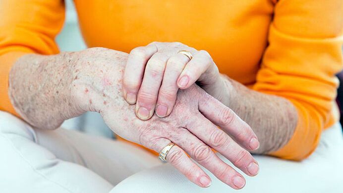
The first symptom of the disease is usually a small joint pain (arthralgia), which bears the biggest burden.This is, first and foremost, the lower-wrot joints, hips, plus-phalanx joints from the first thumb.From the upper limb joints, interphalngeal joints, carpal-shaped brush joints are more often affected.
Arthrosis usually begins with a wound of one joint, but after several other joints are involved in the process.
The main symptom of arthrosis
With arthrosis, the patient complains of pain, crisis, movement restrictions in the joints, swelling and joint deformation.
Separately, it should be experienced in pain.With arthrosis, mechanical and startup pain is possible.Mechanical pain occurs with a load on the affected joints.Such pain is very difficult in the evening at rest, disappearing after a few hours of rest.The appearance of this type of pain is associated with gradually increasing bone pressure during physical work.Pressure causes bone beams and painful bone tissue irritation.
Start the pain appears at the beginning of the walk, then stop quickly and occur again during the physical work.Starting pain can appear with the articular surface friction of the affected joints.Small particles of necrotic cartilage fall on the surface of the cartilage.In the first step, these particles are pushed into the joint bag cavity and the pain stops.
With arthrosis, pain can be associated with periarthritis and tendource (soft periartic tissue inflammation, ligaments and articular bags).This pain occurs only during movement in which the affected tendons participate, as well as in certain positions of the joints during movement.
Pathological changes, as a rule, begin with large joints, subject to large physical work during the day.At the beginning of the disease, pain occurs as a result of the inconsistencies of the micro channel with the need for articular tissue.Therefore, to reduce the pain, the patient slowly takes the first few steps and only accelerates the rate of walking.Pain can appear after half to two hours of walking or working in a standing position.This is a signal for changing loads, short breaks or types of work.
In the final stages of the disease, arthralgia can occur with a minimum burden on the joints and remain relaxed for a long time.This is due to the fact that in later stages, rough changes in the joint tissue, the destruction of the articular cartilage, and secondary synovitis are formed.With the major development -the rough changes in the bone tissue, the individual fragments can be separated and, falling into the joint gap, causing sharp pain.This phenomenon is called the symptoms of mouse together.
During joint examination, deformation should be noted.In addition, with arthrosis, there is a thickness of periastal soft tissue, regional muscle hypotophy, limb axis displacement.Interphalangeal joint thickening with bone growth and periarticular cloth seal is called gerberden node.
Pain when feeling joints is localized in a joint gap, places of joint capsule attachment, but the symptoms of the disease are not always.Swelling and joint pain are determined by secondary synovitis.
Violation of mutual function in the early stages of arthrosis is indicated by the limitations of the amplitude of the movement.This is caused by periosematic tissue and synovitis.
In the final stages of the disease, the clinical manifestations of the contract develop vary in terms of severity.Often, the function of the knee and hip joints is affected.
Symptoms of arthrosis depend on pathological localization
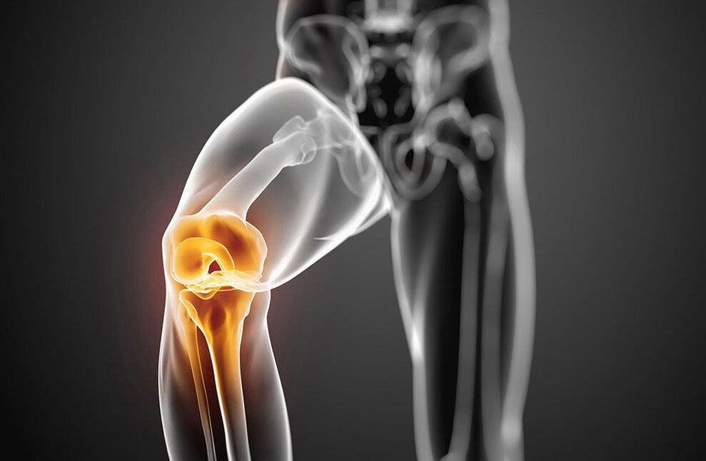
Arthrosis with damage to the knee joint - symptoms
Knee joint wounds with arthrosis are called gonarthrosis.The main gonarthrosis develops in women in menopause.The secondary causes are the most frequent knee joint injury and statistical violations with spinal curvature, flat feet.The patient complains of pain in the knee joint that occurs during movement, especially when walking up the stairs.The pain is localized in the front or in the knee joint.The movement in the joints is limited: the first bending, and then the extension.When moving, problems often arise.With the development of reactive synovitis, pain during movement is increasing and resting anxiety.Swelling of the joints, pain during palpation, redness (hyperemia) and increased skin temperature are determined.Over time, due to bone growth, knee joint deformation occurs.
Arthrosis with damage to the hip joint - symptoms
The hip joint wound is called coksartrosis.This is the worst form of arthrosis.Causes of this disease may be congenital displacement of the hip joint, injury, menopause.The patient suffers from joint pain during movement, in a standing position.Movement restrictions in the joints gradually increase (the first internal and external rotation, Flexion later).There are flaws related to shortening the limbs.With bilateral damage, duck gait is typical.Muscle atrophy -The thighs and buttocks develop.No joint swelling with cartroses.Palpation determines limited pain in the femoral head.
In the early stages of arthrosis, joint functions are preserved.With further development of the disease, it starts to be temporarily limited, and then the ability to work completely, patients lose their own ability, need help outside.
The cause of arthrosis
Arthrosis is based on the main degeneration of the articular cartilage with damaging changes that are included in the bone that form the joints.Such degeneration occurs due to the imbalance between the mechanical load on the surface of the cartilage and the possibility of compensation for this load.
In the development of degenerative changes in articular cartilage, several factors can simultaneously participate:
- Functional advantages, including professional, household and sports, cause mycotrauma cartilage;
- joint injury;
- infectious and non -specific joints;
- joint displacements, leading to violations of joint surface comparison;
- Violations of the body's statistical due to spinal curvature (kyphosis, scoliosis, pathological lordosis, etc. -other), flat feet;
- Chronic hemarthrosis:
- diseases with metabolic disorders (gout, obesity, chondrocalcinosis);
- Osteodistrophy or pedgetic disease;
- osteomyelitis;
- Pathology of the peripheral nervous system with loss of sensitivity;
- Endocrine pathology (acromegaly, diabetes, amenorrhea, hyperthyroidism);
- Descent tendency.
The risk factors of arthrosis include old age, female sex, obesity.
The mechanism of development
Metabolic disorders in the cartilage are based on quantitative and qualitative changes in the main ingredient of cartilage.The main ingredient is made up of proteoglican that provides collagen stability.The development of arthrosis is accompanied by inadequate formation or increased destruction of cartilage components.
With osteoarthritis in cartilage tissue, hyaluronic acid content, chondroitin and keratin decrease.In addition, changed proteoglican loses the ability to maintain water.It is absorbed by swollen collagen, causing a decrease in cartilage resistance.
If chondrocytes are damaged, they begin to produce collagen and proteoglycans are not characteristic of ordinary cartilage tissue.These modified ingredients cause loss of cartilage biochemical quality.
What is very important in the development of arthrosis is immune disorders.The destruction of cartilage is accompanied by the appearance of cellular and humoral immune reactions.On the other hand, this causes progressive fibrosis and synovial membrane sclerosis, pathological changes in intraarticular synovial fluid, and cartilage violations.Lower synovial shells support the development of degenerative changes in joint cartilage.
Descendants have certain values in the development of arthrosis.
Classification of arthrosis
Arthrosis is divided into two groups: primary and secondary.
In distribution (main arthrosis):
- Local (with damage to three joints)
- Ordinary or general, polyarthrosis (defeat of three or more joints).
Depending on the destination (secondary):
- A. Tasobed joints (chocolate);
- A. Knee joints (gonartrosis);
- A. elbow joints;
- A. shoulder joints;
- A. spine;
- A. Cervical department (unkoarthrosis);
- A. hands;
- A. Together ankle (Cruzartrosis)
- A. Stop.
By etiology:
- Post -traumatic
- metabolic
- Because of endocrine pathology.
Diagnosis of arthrosis
Various clinical manifestations and variants of arthrosis make it difficult for the early diagnosis of the disease.Diagnosis of falsehood is also associated with a lack of certain symptoms, the onset of hidden disease.What is very important is the definition of factors contributing to the development of arthrosis:
- chronic joint trauma;
- the length of the stereotype movement;
- physical activity at the joints for a certain time;
- violation of salt or fat metabolism;
- The frustration of the descendants of the musculoskeletal system.
The X -ray examination is the most important meaning in the diagnosis of arthrosis.Radiography of the second view -two knee joints are performed in a direct position, a crooked position, apart from the side position.Signs of classical arthrosis of radiography are: narrowing of joint gaps, presence of osteophytes, subcondral bone sclerosis and subcondral cyst.There is a stage of radiological changes in arthrosis:
- 0 - nothing changes.
- I - Signs - Doubtful radiological signs.
- II - Minimal changes (slight narrowing of joint gaps, subsidiary osteosclerosis, single osteophytes).
- III - Moderate manifestations (moderate narrowing of the charter, various osteophytes).
- Iv- Expressing changes (invisible joint gaps, various gross osteophytes are determined), synovitis is often present.
In front of these symptoms, further tools are not required.
In their absence or low severity, joints, MRI, scintigraphy are performed.
Clinical trials of blood, urine and intraarticular synovial fluid are not included in the list of mandatory studies for arthrosis diagnosis.But this test is required to exclude the articular pathology.
Signs of clinical arthrosis and main diagnostics:
- Mechanical joint pain;
- fatigue;
- feelings of instability in the lower limbs;
- damage to the first joints of the legs and hands;
- the onset of the disease gradually;
- Slow progressive current;
- deformation together;
- regional muscle hypotophy;
- recurrent synovitis;
- Movement restrictions in joints;
- X -ray changed.
Arthrosis must be distinguished by damage to the joints with rheumatoid arthritis, infectious, metabolic and reactive arthritis.
Rheumatoid arthritis, unlike arthrosis, begins with inflammation of the small joints of the hands and feet.It is characterized by intense pain from the type of inflammation, morning stiffness of the joint, the presence of rheumatoid nodules.
Gotric arthritis is found primarily in men.High local activities with acute paroxysmal pain in the first Plus-Phalanx joints of the toes are characteristic.With gout, the presence of tofus is typical, in radiography there is a "punch".
Psoriatic arthritis is characterized by skin lesions, especially scalp, finger -shaped spindle deformation and bright raspberry color over the affected joints.
Infectious arthritis is characterized by acute onset, rapid development and course, sharp pain, high temperature and the effectiveness of antibacterial therapy.
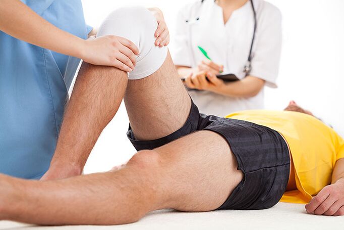
Arthrosis treatment
Treatment for arthrosis should be long, complex.Basic principles of arthrosis treatment:
- Unloading joints (proper mobility mode and mechanical load, walking, weight loss, prolonged position exemption, weight wear, strengthening muscle-ligaments using physiotherapy, massage, electrical stimulation).
- Conservative correction of static disorders (use of orthopedic shoes, corsets, supervisors).
- Effects on metabolism and overall blood circulation (biostimulan use, vasodilating drugs, balneotherapy and physiotherapy courses twice a year).
- Removal of reactive synovitis, anti -inflammatory therapy.
Arthrosis patients show diet with salt, sugar, strong tea, coffee, smoked meat, sharp dishes.This improves the sensitivity of vascular and articular receptors, restores blood vessels, normalizes the exchange in condo.With arthrosis, you need to drink enough fluid (at least 8 glasses of water a day).
Arthrosis drug treatment includes the use of anti -inflammation and rapid painkillers (anti -anti -Non -Ssteroids -NSAIDs), Basic drugs -Chondroprotectors.No-?TSO-2 inhibitors are not selective and selective used from NSAIDs.
As a local therapy for affected joints, NSAIDs in the form of ointment or gel are used.
In the presence of reactive synovitis, tendinitis or tendovaginitis, when NSAID treatment is ineffective, appropriate intraarticular or intramuscular administration.
Basic therapies with chondroprotectors (chondroitin, glucosamine, hyaluronic acid) are used to prevent cartilage degeneration.
Treatment of chondroprotectors is indicated in clinical and radiological stages of arthrosis I-III.
In addition to direct chondroprotectors, drugs that stimulate cartilage tissue recovery (biogenic stimulation) are used.These drugs are used during forgiveness, without the presence of reactive synovitis.
With arthrosis, drugs that improve micro circulation are also indicated.With the presence of varicose veins in the lower leg, correction of venous blood flow is required.
In patients with arthrosis, it is necessary to diagnose and treat timely osteoporosis.
Arthrosis physiotherapy
Physical treatment methods are also related to basic arthrosis therapy.Under their influence, metabolic processes, blood circulation and tissue fluid are stimulated, neurogumoral regulations are restored.
Treatment complexes with arthrosis include inductormia, microwave therapy, pulsating current, drug electrophoresis and magnetotherapy.To eliminate synovitis, ultraviolet irradiation of the affected joint area in the dosage of erythema, the electric field of ultra -high frequency, electrophoresis with analgin, dimexide or hydrocortisone is used.
For the prevention of arthrosis, it is recommended to lose weight, prevent increased load on joints, walking in oppressed areas, increased moisture and hypothermia.The selection of individual shoes and supervisors is important.
With gonarthrosis, normal physical exercise, swimming, cycling is shown to strengthen the muscles.Classes -heavy and lightweight sports, football is not recommended.
Therapeutic exercises are conducted differently, in sitting, lying, in the pool.Movement cannot be intense, traumatic, the volume and number of repetitions increases gradually, avoiding the load.
Popular and effective methods for treating arthrosis also include massage and kinesitherapy.
With significant changes in joints with deformation, mobility restrictions, surgical treatment is recommended.Arthroplasty, endoprosthetics, osteotomy dilakukan.
The prognosis of the disease
Main arthrosis rarely brings complete defects.With the presence of reactive synovitis, patients temporarily become disabled, and sometimes they have to change the profession.With the secondary kokarchy, the prognosis is poor due to the rapid disease of the disease with the development of significant joint function.In such cases, defects can occur for several years of disease.
Prevention of arthrosis
The main prevention of arthrosis should begin in the childhood.It is as follows:
- prevention and treatment of scoliosis;
- Flat foot correction using a special supervisor;
- Physical Education classes to strengthen muscles and ligaments;
- rational nutrition and the prevention of metabolic disorders;
- heavy sports restrictions in children and adolescents;
- Work to sit at the table on a walk;
- Proper labor organization and other employees in the company where there is heavy physical activity.
Secondary prevention provides steps that prevent the development of recurrent reactive synovitis.These include walking, physical work restrictions, walking with support and other steps that unload joints.With severe symptoms of arthrosis, it should always take basic medication.General reinforcement therapy, increased blood circulation and metabolism, annual spa treatment is recommended.
Which doctor will he go?
- Rheumatologist
- Orthopedist